Not all back pain has a definitive cause

“Findings such as disk height loss and disc bulges are coming in individuals without low back pain.”

Disc bulges, degenerative joint disease, spinal stenosis, do you all a result of living in this world. We have gravity acting a force on us almost 16 hours a day. Anytime that there is a problem, we want to blame something or somebody. Low back pain is an enigma at times. we can draw correlations, we can come up with risk factors, we can even tell you how to treat it sometimes, but what we can’t do is tell you exactly what causes your back pain.

“Surprisingly, disc protrusions were associated with a lower risk of subsequent back pain. Nerve root contact and central stenosis had the largest hazard ratios on baseline imaging findings, and they were associated with incident back pain in the expected direction but not statistically significant. Self identified Depression was the strongest predictor of subsequent back pain, with a greeter hazard ratio than any imaging findings.”

What should be taken from the above statistics is that mental health plays a role in pain. There are a lot of new studies that are associating catastrophizing and external locus of control with increased pain levels. Work by Nadine Foster demonstrates screen for patients who will have a difficult time improving with therapy alone. New were books, such as the one by Annie O’Connor and Melissa Kolski (two people with whom I’ve studied at our RIC), goes into great detail regarding pain science. Big picture, we can not neglect the patient’s emotional well-being when attempting to treat the patients physical complaints.

“Our results indicate that depression is a strong predictor of who will subsequently reports low back pain then baseline imaging findings.Subjects with self reported depression at baseline were 2.3 times is likely to have back pain compared with those who do not report depression.”

There is obviously a psycho social component to low back pain. The question is… Chicken or the egg. Is a person more likely to be depressed because they have back pain that is not improving? Or is that person more likely to have back pain because they are depressed? I don’t think that there are cause and affect articles in the literature at this point, but there is definitely a high correlation between patients who are depressed and patient who continue to report low back pain.

“In our analysis of baseline data, we concluded that central stenosis, nerve root contact, and disc extrusion were the most important imaging findings related to prior low back pain. Our current analysis indicates that central stenosis, disc extrusion, and route contact may also be risk factors for future low back pain.”

In other words, if you have a major deformity you will probably have pain. This doesn’t mean that you will definitely have pain, it just increases your risk of experiencing symptoms.

The moral of the story is that we cannot deny the brain. The brain has the ability to see pain, and some patients are more susceptible to seeing this pain. Don’t get me wrong, a thorough mechanical evaluation should be performed when a patient has pain, but when this patient is not inclined to respond to mechanical therapy, the patient should be referred to someone that can better handle this patient’s pain.Sometimes, that person will be a behavioral therapist, a psychotherapist, or a clinical psychologist. Physical therapists are not always the go to in order to treat a patient’s pain.
Excerpts from:

Jarvik JG, Haegerty PJ, Boyko EJ. Three-Year Incidence of Low Back Pain in an Initially Asymptomatic Cohort. Spine. 2005;30(13):1541-1548.

Brother my Brother

Today’s blog is very different from any of those written before. This is an insight into my life, into my thought process, into my experiences, into those things that made me who I am. I started this blog to teach people about healthcare, but there is so many more things that people can learn from my experiences. I dictate today’s blog on my way to the cemetery. It’s a little bit more emotional than anything that I would typically write. 
 Life is precious. My brother is a fucking idiot, in 2008 he overdosed. He never really saw anything outside of Joliet Illinois. I want to live until I die. There is too much to see and too much to live for in this life. After eight years, I still think of everything that he missed out on. He missed out on having a family. My family is the greatest thing that ever happened to me. I would be my fathers son, I would be content to stay at home and work hard and live my life in that fashion. My wife loves to travel and loves try new things. If it wasn’t for her I would’ve never traveled to Europe, I may have never made to Alaska. If it wasn’t for my daughter, I wouldn’t slow down and slide down the big slide. I probably wouldn’t go to another waterpark, I probably wouldn’t climb in the tunnels at Odyssey fun world. My brother missed out on a lot, when I go visit cemetery it just my heart.. Life is precious. For those going through difficult times, Know that life is precious. There are people that love you and people that will miss you if you’re gone. I miss my brother frequently. Life goes on, and life will go on without you. I hate to say that because it sounds harsh but it will. I am happy, and unfortunately he’s not here to see that. 

At what age does dreams die? I don’t know that answer. At what age do we throw in the towel? At what age do we give up? I don’t know what was going through my brothers head those last days And it kills me eight years later. 
In memory of Michael Anderson. I miss you brother

Does taping in addition to PT provide increased benefits?


This is a look at a popular form of taping using in the PT profession. This was popularized in the Summer Olympics years ago and has increased in usage in the PT profession, regardless of what the evidence states.


  1. “Low back pain is a significant public health problem that affects approximately 39% of individuals worldwide at some point in their lifetime”


This is like beating a drum. If you follow the blog, I have written many times over the year regarding how expensive back pain is in the developed countries. One aspect that surprises me is how low this number actually is. In other articles, it talks about the lifetime prevalence rate between 70-80%. I would have to surmise that “worldwide” changes this number. I don’t have the reason why, but I have my guesses. I would guess that those “undeveloped” countries are spending less time on their kiester and more time either in a deep squat or standing position.


  1. “Several interventions commonly used by physical therapists, such as manual therapy techniques and exercises, are endorsed in most guidelines as effective treatments for patients with low back pain…”


Moving is better than not moving (in most cases). It’s funny because when I was a personal trainer (many, many years ago) I used to think of Physical Therapists as overpaid personal trainers. I completely disagree…sometimes. Don’t get me wrong, there are some PT’s that only prescribe 3 sets of 10 repetitions because it is traditional and for those PT’s I would agree that they are overpaid personal trainers. When prescribing exercise, we always have to think; “what’s the goal”. If the goal is pain reduction, than 3 sets of 10 may not be appropriate. If the goal is absolute strength or power or endurance, then 3 sets of 10 may not be appropriate. If the goal is hypertrophy…you got me…it may be appropriate for some patients for some muscle groups. In the end, 3 sets of 10 for everyone is no better than 3 sets of 5.


This isn’t meant to blast the PT profession, but if you are being treated in PT…Look around! If you are doing the same exercises as everyone else, then you have to question whether you are exactly like everyone else?


  1. “Kinesio Taping method was introduced at the Olympic Games in Athens and has since gained in popularity”


We have seen these tapes for the most part. The colorful tape worn on shoulders or backs of athletes. In the summer games, especially for women’s volleyball (I’m sure other sports have them, I just seem to watch more of this than anything else except for weightlifting), these colorful tapes are apparent. I use the tape, not for the reason indicated, but it makes for a great thumb wrap when using the hook grip in weightlifting.


  1. “The evidence of the benefits that Kinesio Taping can provide for patients with chronic low back pain is still scarce”


I could sell a cup of water to a drowning person in the ocean. I could easily sell Kinesio taping to my patients and others in the athletic arena, but I have yet to read a well-performed study that shows it is better than not using Kinesio tape. It’s the modern day ultrasound…It works until it doesn’t.


  1. “There is no current evidence to support the use of this method.”


This is not to say that it doesn’t work…yet, but of the studies performed thus far…it doesn’t work. One of two things will happen over time: 1. The company(ies) that sell the tape will continue to publish their own case studies to show the efficacy and/or 2. The peer reviewed journals will stop publishing all of the negative studies because academia will stop performing studies that consistently give the same results.


  1. “…the objective of this randomized controlled trial was to compare the effectiveness of adding Kinesio Taping to a physical therapy program in patients with chronic nonspecific low back pain.”


This is a well-performed study. Randomized doesn’t mean that the study is done randomly or half-assed, but the people in the study (guinea pigs) are separated in a scientific manner.


6a. Misc: There is a bunch of instructions for how the study was actually performed in the Methods. This is boring to the non-medical reader, and sometimes boring for those of us that read research. I will spare you the details. Just know that the study is well-performed.


  1. “The group that received physical therapy plus Kinesio Taping had the elastic tape applied to the lower back at the end of the sessions”


Essentially, if the tape is to provide greater benefit than exercise alone, this group should outperform the exercise-alone group in the data measured.


  1. “The corresponding author is certified by the Kinesio Taping Association International and provided training to the therapists on how to apply the Kinesio Tape”


This is important. If there is a method to perform on a patient, but the participating therapists are not certified in the method, then it could be that the practitioner doesn’t know the method well enough to perform the method. Since at least one of the authors is certified, it would make this a moot point.


  1. “After 5 weeks of treatment, the between-group comparisons showed no advantage of using Kinesio Taping in these patients for all primary outcomes…the addition of Kinesio Taping to physical therapy did not enhance treatment outcomes at any point in time.”


Crickets chirping………….Enough said.

  1. “Our data corroborate the results of 3 previous randomized controlled trials that do not support the application of Kinesio Taping in patients with chronic nonspecific low back pain.”


This means that if you want to tape your thumbs in order to lift weights, then go ahead, but using this type of tape (there are many different manufacturers of this type of tape) for back pain may not be ideal.


QUOTES TAKEN FROM: (Also, the initials of the first author is actually MAN, that’s awesome)


Added MAN, Costa LOP, De Freitas DG, et al. Kinesio Taping Does Not Provide Additional Benefits in Patients With Chronic Low Back Pain Who Receive Exercise and Manual Therapy: A Randomized Controlled Trial. J Orthop Sports Phys Ther. 2016;46(7):506-513.

Slump test: what’s it mean?

This is a common test performed in the clinic. The article goes into great detail (again not written about here because I don’t really have an opinion about the technique) of how to perform the test. I highly recommend reading the article for all health care practitioners that care for patients with spinal pain.


  1. “neuropathic pain (NeP) as “pain caused by a lesion or disease of the somatosensory system”


This is a great start to what will be a good read. If I were to say this to most PT students that come into my clinic, I would get the “Bambi in the head light look”. The fastest way to say this is that neuropathic pain is a pain that may be coming from a structure that is innervated (has nerves). This doesn’t really tell me anything though.


  1. the presence of NeP has been linked with poor recovery, along with higher health care costs and lower quality of life.”


This makes sense to me. If you have pain, that is coming from somewhere, you are more likely to require more treatment than someone that has pain coming from nowhere and are less likely to enjoy your life than someone that is pain-free.


  1. “The diagnosis of NeP typically consists of a thorough history and an extensive neurosensory examination to identify both positive (exaggerated responses to stimuation, such as allodynia) and negative (various sensory and motor losses) signs…usually performed by a specialist, requires a lot of time to complete, and in many regions involves a long waiting period for the consultation”


As a Doctor of Physical Therapy, I happen to be said specialist. This type of patient typically takes longer to evaluate than someone that only consists of weakness or deconditioning. There is much more to look at. I don’t know if I would say that it involves a long waiting period for the consultation. I think that this has more to do with a person’s type of insurance regarding wait time. For instance, if you are willing to pay out of pocket for the assessment, and pay what the “chargemaster” (an inflated charge board that no one ever really pays…unless they can’t afford insurance) states, then I am sure that I can fit you in for a $400-600 evaluation tomorrow. (We don’t actually make that much for an evaluation, but insurance companies would love you to believe that we do).


  1. “The straight leg raise (SLR) test is the most commonly used neurodynamic test for the lower extremity. The slump test is another…”


The SLR test is easy to perform and most physicians are aware of its implication in discogenic pain. I include it when I am trying to make a point to physicians and insurance companies regarding a possible pain generator. I prefer to use the slump test because it is easier for me to test when I already have the patient sitting in the chair testing lower extremity strength (testing nerve electrical power). If I have a positive slump test, then I will typically perform a SLR test in order to “paint a better picture” of the patient to the insurance company.


  1. “the SLR test demonstrated 100% specificity in patients clinically diagnosed with NeP.”

I don’t expect the lay person to understand this because this is such a difficult concept for most physical therapy students to understand. Basically, if you have a positive test, then your symptoms may be coming from the nerve. If you have a negative test, then we can effectively rule out nervous tissue pain.


  1. “Key components of the clinical exam included pain (location, behavior, quality), motor function (strength/weakness, reflexes), sensory function (mechanical/thermal sensation), autonomic function (sweating, hypotension), and the SLR test”


You’d be surprised (or may you wouldn’t if this also describes you) regarding how many patients are surprised that a PT would take their blood pressure. I read a statistic years back regarding the following: 50% of patients seen in an outpatient clinic have undocumented hypertension. I can’t tell you how many times I have had to stop a session to call a doctor to inform them of a patient’s hypertension. I had one patient argue with me years ago that he wasn’t there for blood pressure issues, but to have his pain fixed. Unfortunately, his BP was higher than the maximal allowable threshold for exercise and I sent him back to the doctor. (Mind you, my current state is not a direct access state, which means that the patient had to be referred by someone prior to coming to therapy). The patient was apologetic the next session when the doctor told him what the numbers could mean regarding DEATH! That’s right…I save lives. Just kidding, but not really. If you see a therapist, your blood pressure should be checked at least on the first visit. If it is not, question the intentions/ignorance of the therapist. Could be oversight on the therapist’s part. I used to be that therapist.


Also, your sensation and strength should be tested in certain circumstances. Again, just ask your therapist why/why not the above testing was/wasn’t completed. There should be a good rationale for the answer. We love to teach…at least I do.


  1. The authors of the article did an excellent job of describing positioning for testing, so if you are in health care and would use the slump test, it is a good refresher. Highly recommend it.


  1. “…designated as positive if the following conditions were met: (1) pain or sensations were reduced with neck extension, and (2) there was a right-to-left difference in pain distribution or there was a difference between right and left knee extension.”


This is something that most PT students, and I must surmise that new professionals miss while in the clinic. This is the small details of the test. Is there a loss of knee extension on one knee? The only way to know is to have someone teach it to the student or for the student to go out on his/her own to learn it. What profession are you in? In your profession, who is the best in your workplace? What separates that person from anyone else in the workplace? Usually it is initiative, persistence, self-learning, confidence, experience, etc. This same thing can be said for the PT profession. We aren’t all born with these characteristics; they are honed over years of working and studying. If all we do is work, then we may gain experience, but the experience may be that of missed details.


  1. “From these data, a difference of 10 degrees or more was used to indicate a positive slump test component.”


Again, small details. Many students are not looking at the angle change in the slump test and although 10 degrees is small when eye-balled, it can be huge when it is the difference between being able to stretch out and push the gas pedal with or without pain.


  1. “very little can be interpreted when the slump test is positive but the pain does not extend below the knee.”


Students need to know this stuff! We learn in school that if it produces pain, that the test is positive, especially if the pain is reduced when the patient is then asked to look up. This means little in terms of telling us important information.


  1. “The sequential combination of the 2 tests provided an effective means of ruling out those without NeP and ruling in a large proportion of those with NeP.”


This is important for students and PT’s to understand. If the patient has no pain production with the slump test, then “nerve pain” can effectively be ruled out due to the small rate of false negatives. When a patient has pain below the knee, then “nerve pain” can be ruled in.


This was an excellent article going more in depth than anything we learn in school. I have been using this test for years and have slowly incorporated the information into practice over the years, but I now know that there is a good reason to look at the knee extension change. When studying the McKenzie Method (MDT), we are taught to look at the angle changes when performing these “dural tension” based tests. It is informal when taught in the course, but here is the formal information.


Quotes taken from:


Urban LM. Macneil BJ. Diagnostic Accuracy of the Slump Test for Identifying Neuropathic Pain in the Lower Limb. J Orthop Sports Phys Ther. 2015;45(8):596-603.

Traction: useful or not?

I use traction sparingly. It is a last resort if the patient is going to have a surgery. If I have tried everything in my power and knowledge to help a patient, and the patient continues to not improve, then traction it is. It is my Hail Mary.

  1. “Physical therapists may choose from myriad intervention options for LBP, but the effectivenss of many of these options is questionable”

Do you feel good about coming to therapy yet? An awesome question to ask your therapists is; “What does the research say about xyz?” or better yet “Does the research support xyz for my condition?”

It’s funny, in school we all learn that ultrasound brings more blood flow to an area…SO FRIGGIN WHAT? Does that blood flow actually fix me? Not really, but it brings more blood flow! That’s an expensive transportation of blood. Do you know what else brings more blood flow…Hickies. That brings more blood flow. Ask your therapists to suck on your skin for a while to see if that will also bring more blood flow. It will probably cost you a little more for that service though…I digress. There is not much, if any, CURRENT research that supports the use of ultrasound for back pain. If your therapist tells you that ultrasound will help, ask how? If they tell you the blood flow thing…ask them to pucker up.

  1. “Authorities have recommended traction for conditions including protruding Intervertebral discs, spinal muscle spasm, and general pain and stiffness”

This is what I learned when I was in school. Seems archaic that we were taking general recommendations from “authorities” to try to fix the second largest complaint to the common cold. At least research has advanced from the opinion of authorities.

  1. “several systematic reviews and clinical guidelines conclude that the effectiveness of traction is limited…little evidence to recommend traction…clinically important benefits of lumbar traction were demonstrated for neither acute nor chronic back LBP…traction should not be used…41% of the physical therapists in the UK used traction”

Boy can those statements be any stronger. Traction should not be used because it is not very effective for low back pain (LBP). Now if you want to use traction because it makes you feel better, then go ahead. Sugar pills work for some people also. (Not trying to come across as sarcastic, but I’m sure it sounds that way). If you have preference for a specific intervention, then that intervention may be likely to help you. I have a patient that believes that ultrasound of the back muscles helps. No matter how much education I have provided, I’d be better off talking to the wall. Needless to say, we have a great conversation during the ultrasound, while the patient is propped up on his elbows and lying on his belly. For those that know, these positions can help/fix up to 65% of back pain patients.

  1. “Our findings suggest that a majority of APTA Orthopaedic Section members use traction…In contrast, approximately one third of respondents indicated that they would use tractions for patients in a manner that is contrary to that classification”

There is a clinical prediction rule in the derivation (creation) phase that indicates a certain type of patient may benefit from traction. This is less than 10% of the patients in the clinic. This rule has not been replicated yet, so it is more like an educated guess at this point. Other research has reported the above, in which traction has no added benefit to an exercise program. Also, exercise increases blood flow (see above). The sad part is that about 1 in 3 therapists are using traction contrary to how it should be used. Have you seen that therapist? They are typically the ones applying hot packs, Hickies and massage.

  1. “employing soft tissue mobilization or massage was identified by approximately 65% of our respondents as a supplement to traction. Given limited evidence for the effectiveness of massage for treating LBP…the extent to which physical therapists in the United States use soft tissue mobilizations/massage in managing LBP may be concerning”

WOW! I was totally talking out of my a$$ in the above paragraph, but my a$$ is also supported by research. Who knew?

  1. “there is a growing body of evidence that higher levels of professional preparation influence clinical decision making and, potentially, patient outcomes”

Look there has been a backlash in our profession for what is called “alphabet soup” after our names. This means that some therapists have gone on for “extra” training and certifications. This is important. Unfortunately, our profession has deemed it inappropriate to put down all of the certifications after our name. The only way to know what your therapist knows is to ask. I personally have the initials:

DPT (Doctor of Physical Therapy), cert. MDT (certified in Mechanical Diagnosis and Therapy). None of the above initials were given to me…I earned them.

Thanks for reading this. If I go overboard at times and offend you, there are other blogs to read. Have a good night.

Quotes taken from:

Madson TJ, Hollman JH. Lumbar Traction for Managing Low Back Pain: A survey of Physical Therapists in the United States. J Orthop Phys Ther. 2015;45(8):586-595.

Soft sell to patients

Soft sell to patients


  1. “Your clients do not know what you know”


Man…this statement says a lot! We are highly educated (some more than others of course) and our patients come into the session with varying levels of education regarding either their health or the specific ailment. The part that irks me though is when the patient DOES know more than my students. We dedicate so much time to teaching our patients that I am frustrated if my patient now knows more than the student working with the patient.


Don’t be offended, but I am going to talk to you like an 8 year old, until you’ve earned the right for me to talk to you like a teenager. I will talk to you like a teenager until you’ve earned the right to be spoken to like a college student and so on and so forth. I have to ensure that you know what I am trying to teach you. If that means that I have to dumb it down a little at first…so be it. Senor Sosnowski once said that a smart person can always climb down the ladder of intelligence, but an ignorant person can’t just climb up the ladder. They have to put the work in order to get to a level of intelligence on this topic. I will be the first to say that I suck at a lot of things…physical therapy just isn’t one of them.


  1. “simply calls for a direct and simple correlation that is made between your intervention and the positive outcome achieved by your patient.”


I expect people to improve. With patients that I don’t expect to improve, I am over educating that patient on day one. This is few and far between though. I expect patients to improve and in the end, I will never act the hero, but more like the facilitator. When you understand that you are “in charge” of your symptoms, then I become your cheerleader. (I’ve worn heels, but won’t go so far as to wear the skirt…one day I’ll tell the story of the heels).


QUOTES TAKEN FROM:


Quatre T. Why they buy: Because You Have connected the Dots. IMPACT June 2016:11.


MRI’s fact vs fiction


This was a great article. It puts numbers to the faces seen on MRI’s. I like numbers…kind of like Rainman. Numbers comfort me. Enjoy the read. There is some higher level thinking in the below quotes. If you have any questions, leave a post either here or on the movementthinker Facebook page.


  1. “Magnetic resonance imaging (MRI) provides clinicians with a noninvasive mechanism for viewing lumbar anatomy in great detail”


READ AND RE-READ THE ABOVE STATEMENT.

Question #1 from the above statement: Can an MRI tell me what is causing your pain?


Question #2: Can an MRI tell me how to treat you?


Question #3. Does the MRI differentiate between abnormal structures that cause pain and abnormal structures that don’t cause pain?


The answer to all of the above questions is NO! Everyone seems to think that they need an MRI before they come to therapy…as if I am going to just treat them on a whim without the MRI…or that the MRI will somehow give me a paint by number way of treating the symptoms. This does not exist. The MRI can be helpful in a small percentage of patients that are either seeking or needing surgery, but aside from that it is just something for me to read after I have performed my clinical assessment of the patient and come up with my own conclusion. Now…if my conclusion matches the MRI then awesome! Well…at least for me. If it doesn’t match the MRI…that sucks because now I have to go back and reassess to see which one of us is more right…the PT or the MRI.


  1. “For example, large variations in lumbar disc and radicular canal morphology have been identified in both symptomatic and asymptomatic individuals”


This means that an MRI is very good at determining what is not normal, as compared to a textbook, but the variations of normal is so wide that the test may not tell us much.


  1. “…challenge for examiners in their attempts to differentiate between observations that are “symptom generators” and those that are benign variations”.


When a radiologist reads your MRI, they are the ones that are determining what is going on in the pictures, they spend on average of 30 seconds per picture. In 30 seconds, they have to figure out what is abnormal. Then, if they have found something abnormal, they have to determine if it can cause your symptoms. All of this is performed without ever evaluating the most important aspect of the symptoms…YOU! The radiologist never sees you. If you look at the bottom of your report (assuming that you have already had an MRI), you will typically see the phrase “patient would benefit from clinical examination to correlate imaging”. This is the radiologist saying; “Look, I only have the pictures. I can tell you with a degree of certainty what the MRI says…does this fit your symptoms?”


  1. Patients were classified according to this table:


  1. Primay LBP (low back pain): pain in the back or buttock


  1. Posterior thigh referral: pain in one or both back/lateral thighs with or without LBP


  1. L1-L3 distribution: pain in the anterior thigh and top of foot


  1. L4-5 distribution: pain in the mid and distal anterior thigh, anterior leg and top of the foot.


  1. S1-S2 distribution: Pain in the lateral border of the foot and bottom of the foot


  1. Bilateral distribution: any combination of the above in both legs instead of one leg.


  1. Atypical: none of the above.


This is an overall pain pattern distribution. Unfortunately, this is not drilled in PT school. I was about 2-3 years out before I figured this out on my own and then after discovering it, I looked it up. It’s funny…if you don’t know what you don’t know, then you don’t know how to find it. I think that PT’s schools should heavily bias students in this direction for learning. Think of it. If you knew that for every dollar you invested, you would get an 80% return if you simply knew a few tricks…would you learn those tricks?


Roughly 80% of the population will have back pain at some point in his/her life. This is either the primary or secondary reason for physician office visits (depending on which research you read) and the one that it competes with is the common cold. Think about that…back pain is about as “common” as the cold.


  1. “All images were initially screened for evidence of neoplastic, inflammatory or infectious disorders…”


This is all of the very bad stuff that needs to be ruled out if someone is going to look at an MRI. This is stuff that won’t get better with therapy. If you have certain characteristics, your PT may refer you back to your physician in order to rule out the nasty stuff.


  1. “…study involved 408 participants…55% had acute pain…50 participants reported a recurrence of previous symptoms within the past 2 months…303 participants reported chronic symptoms of longer duration than 2 months”


This sounds about right. Those with back pain may have it go away, but it will come back. Those whose pain doesn’t come back is mostly because…IT NEVER WENT AWAY!


  1. “…the most common location of symptoms was in the S1-S2 segmental, followed by the L4-L5 distribution. Bilateral radicular patterns were the least frequent.”


This means that a high percentage of patients had symptoms radiating into the foot, from the back. Fewer patients experienced symptoms into both legs. If both legs are causing you pain…at the same time…you are among the few.


  1. “The presence of weakness in ore of both lower extremities was reported by 175 participants (42.9%)”


If your back symptoms are bad enough, they will start to cause a “power outage”.   For instance, I use a specific analogy in the clinic. If your lamp doesn’t turn on when you flip the switch…what is wrong?   A common answer is that the light bulb is burned out. How many light bulbs will you go through before you realize that the bulb is working fine? When a muscle is weak, it is like the above idea. I can give you strengthening, but I would have to give you about 6 weeks of strengthening exercises in order to determine if “just muscle weakness” is the problem. This is like changing the light bulb daily for 6 weeks. I doubt that you would actually do this. Most people may do this once or twice and then just give up. When I give you strengthening exercises, you will do them for a couple of days and then give up because you won’t see much change.


What else could cause the light to not turn on? There could be a fray in the cord. This also happens in the body. If there is a nerve (electrical wire) that is not working appropriately, then the muscle won’t contract…the light bulb won’t turn on. This one becomes a little harder to figure out because we would have to try to find the location of the “fray”.


The final thing is the easiest to check for…the lamp isn’t plugged in.


It’s funny because I frequently have students. Recently, I had a patient that struggled to go up the stairs. She noted that her leg was weak. Students always want to make a muscle stronger. They are good at that. Unfortunately, her hip muscle wasn’t plugged in. After performing 30 repetitions of repeated extension in lying, her hip strength went from weak to moderately strong. Her ability to ascend stairs was visibly improved and the patient was surprised that her sensation of strength had improved. The student asked “why don’t we learn this in school?” I don’t know. I have the same question.


  1. “Disc extrusion was significantly related to the presence of distal lower extremity pain…not significantly related to weakness…not significantly associated with the presence of paresthesias or numbness”


What is a disc extrusion? This guy does a great job of explaining it: http://www.bodiempowerment.com/disc-bulge-why-is-my-disc-bulging/

Why reinvent the wheel?


  1. “Overall 149 of the participants (37%) had MRI evidence showing some degree of nerve or thecal sac compression…The most common segmental level of compression was L4-L5, followed by L5-S1…There was a significant association between the side of nerve compression and the side of pain…of the 256 patients with no evidence of nerve compression visible on MRI, 151 (58%) indicated unilateral lower extremity symptoms”


This means that some patients that have an MRI will show that the disc has caused some sort of nerve compression. When this happens, you will typically have pain on the side of the compressed nerve. On the flip side though, you can have pain on in one leg that is not coming from the nerve. Think like this…nerve compression can cause leg pain, but not all leg pain is caused by nerve compression.


  1. “participants who reported weakness had a greater prevalence of nerve compression, and those without weakness had a lower prevalence of nerve compression”


Again, the nerve supplies electricity to the light bulb. If the electricity is not getting there because of a problem with either the plug or the cord, then the muscle won’t work.


  1. “Roughly 63% of the participants had no evidence of nerve root compression on MRI. Of these, 35% had pain patterns referring distally to the knee”


THIS IS HUGE! PT’s in school learn that if you have pain below the knee that there must be some nerve that is compressed. This is not always the case. Any structure that has a nerve going to it can cause pain to radiate in a pattern specific to that nerve. For instance, in the neck we know that if we irritate the nerve in the joint, it could refer pain into the shoulder blade. It doesn’t have to be a “PINCHED NERVE”!


  1. “the presence of disc extrusion or ipsilateral, severe nerve compression at one or multiple sites is strongly associated with distal leg pain. Mild to moderate nerve compression, disc degeneration or bulging and spinal stenosis are not significantly associated with specific pain patterns.”


I enjoy weightlifting. When I see a snatch done well, it is like poetry. I can’t explain the entire movement in one fell swoop other than to say it is beautiful. When I see someone do this movement, with little experience, we can officially say that: yes you went from point A to point B, but not well.


When we see a severe nerve compression or disc extrusion, we can say “YUP I KNOW WHAT THAT IS.” Anything past that is a guess as to what is causing your symptoms, based on the MRI.


Quotes taken from the following:


Beattie PF, Meyers SP, Stratford P et al. Associations Between Patient Report of Symptoms and Anatomic Impairment Visible on Lumbar Magnetic Resonance Imaging. Spine 2000;25:819-828.


Frozen shoulder: when it doesn’t move

frozen-shoulder1Frozen shoulder, when it doesn’t move.


Frozen shoulder is a common diagnosis in the clinic. I have seen this problem treated in so many different ways that some PT’s are able to drive Escalades. The problem is that not all treatments are created equal. Educate yourself on what the problem is and how it can and should be treated. It’s your body…understand it at least.


  1. “Frozen shoulder, or adhesive capsulitis…painful and limited active and passive range of motion…reported to affect 2% to 5% of the general population”


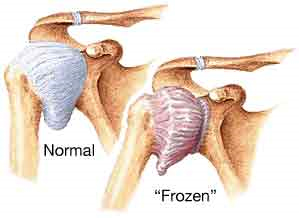
To be frank, frozen shoulder means your shoulder is frozen…it doesn’t move. Adhesive capsulitis is the medical term for…your shoulder doesn’t move! If you take something that does’t move and you try to move it…it is painful. It is not as common as everyone would like to believe and honestly I rarely see it in the clinic. You can have a stiff shoulder and not necessarily have “frozen” shoulder. It affects those that are diabetic more often than those that aren’t, but aside from this, the reason for it is still not certain.


  1. “The absence of standardized nomenclature for frozen shoulder causes confusion in the literature”


We know some things for certain. Your frozen shoulder will go through stages from start to end, what we aren’t certain of is how many stages, and what do we call these stages?


  1. “Secondary frozen shoulder was defined by 3 subcategories: systemic, extrinsic, and intrinsic…secondary frozen shoulder related to insulin-dependent diabetes are more likely to have a more protracted and difficult clinical course”


If you have frozen shoulder because of some other problems, this is classified as secondary. If that problem is due to a body disease, extrinsic is due to an injury outside of the shoulder and intrinsic is a known problem of the shoulder.


  1. “another classification system based on the patient’s irritability level (low, moderate and high) that we (the authors of the journal article) believe is helpful when making clinical decisions regarding rehabilitation intervention…Patients with low irritability have less pain and have capsular end feels with little or no pain; therefore, active and passive motion are equal and disability lower…typically report stiffness rather than pain as a chief complaint…high irritability have significant pain resulting in limited passive motion (due to muscle guarding) and greater disability…pain rather than stiffness…”


This is very easy to follow…walk with me. Your irritability is literally that, when you move how irritating is it? If it is not that painful and you have a capsular end-feel (only to be determined by someone that has moved thousands and thousands of shoulders so that it can be determined if the joint is normal or not very moveable), then it is lower on the scale of irritable. If your shoulder feels like a hot poker stabbing you in the eye and twisted every time you move the shoulder…it’s probably highly irritable.


  1. “recent evidence identifies elevated serum cytokine levels as part of the process. Cytokines and other growth factors facilitate tissue repair and remodeling as part of the inflammatory process…sustained inflammation and fibrosis…although the initial stimulus is unknown.”


This is HUGE, for those that are nerdy regarding physiology. Tendonitis…doesn’t exist. That’s a lie, but not far off. When you think that you have a tendonitis, by the time you see a doctor, it is probably a tendinosis. This means that after a short period of time, there are no longer inflammatory markers (chemical of inflammation) in the tendon. The fact that there is sustained inflammation is…NO GOOD! Think about having constant cycles of inflammation going on in your body. It sounds painful. It is! Others have challenged the premise of adhesive capsulitis, in that the capsule itself doesn’t have the inflammatory markers. At this time, it is semantics, because the shoulder is still painful.


  1. “3 sequential stages: the painful stage, the stiff stage and the recovery stage” others have described “4 stages…the preadhesive stage, the acute adhesive or freezing stage…the fibrotic or frozen stage…the thawing phase” these phases may take 12-18 months and “mild symptoms may persist for years”


Although we can’t fully agree on how many stages and how to describe the stages, we know that this is will take a long time in order to become fully functional.


  1. “A full upper-quarter examination is performed to rule out cervical spine and neurological pathologies”


I can’t stress this enough. Just because your shoulder hurts, doesn’t mean that your shoulder is the problem. I refer to the spine as the great chameleon. It can mimic damn near any symptom that you experience in the periphery. If you don’t fully evaluate the spine…or at least take a quick peek…then you may be treating the wrong thing!


  1. “typically reveals significant limitation of both active and passive elevation, usually less than 120 degrees”


Quick lesson, active elevation is your ability to raise your own arm. Passive elevation is your ability to allow me to raise your arm. Those with rotator cuff tears or issues typically have horrible active elevation, but passive elevation is much better and may be normal.


  1. “Scapular substitution frequently accompanies active shoulder motion…” and “Cyriax described a capsular pattern he believed diagnostic for adhesive capsulitis…it is not consistently seen in patients with frozen shoulder when objectively measured.”


Scapular substituion is elevating the shoulder blade in order to perceive that your are raising your arm further overhead. I tell patients to look at the space between your shoulder and your ear. If there is a huge change in that space when raising your arm overhead, then something is wrong. Patient’s will understand this visual. Have them do it with their “healthy” side so that they can see how much space change actually occurs and then do it with the problematic side to compare.


Cyriax, think Alfred Hitchcock look-alike, is one of the greats that provided many thoughts in the infancy of our profession. His theories are still taught in school and we still have to memorize his paradigms for examinations. In real practice though, we don’t always follow his teachings because…they aren’t always right. Each therapist will learn through seeing thousands and thousands of shoulders, that his patterns aren’t always right, but aren’t always wrong.


  1. “Although authors of textbooks have described patients with frozen shoulder as having normal strength and painless resisted motions…revealed significant weakness of the shoulder internal rotators and elevators.”


In school we learned that frozen shoulder doesn’t affect strength. I am not sure if it is still being taught, but I have to believe so because the boards (think OWL exams from Harry Potter) are based on the text books and not on recent research. Regardless, theses patients do demonstrate weakness. In my opinion, this weakness may be related to disuse due to pain or pain inhibition, but that is a story for another day.


  1. “Significant loss of passive external rotation with the arm at the side, as well as loss of active and passive motion in other planes of movement, differentiates frozen shoulder from other pathologies…Early frozen shoulder may be difficult to differentiate from rotator cuff tendinopathy because motion may be minimally restricted and strength testing may be normal”


Big picture…frozen shoulder will present with multiple losses of motion in many planes. Early frozen shoulder will still have ROM limitations, but not as bad as those that are in the second stage, which may make it difficult to see at first. The therapist/MD may not immediately recognize frozen shoulder and the treatment may be inconsistent with what is needed.


  1. “The definitive treatment for frozen shoulder remains unclear…Establishing treatment effectiveness is also difficult because the majority of patients with frozen shoulder significantly improve in approximately 1 year; therefore, natural history must be considered”


In other words, we think we know how to treat it, but even if we don’t you will get better over time. Is it possible that you don’t need to come to therapy…of course! Will you benefit from therapy…of course! Even if the therapist is providing stuff that doesn’t work…like ultrasound…the therapist should be spending adequate time with you in order to educate you regarding the condition and the overall prognosis. If your health care provider is not doing this…walk away! There are therapists on almost every corner if you look hard enough. Find one of quality.


  1. “Explaining the insidious nature of frozen shoulder allays the patient’s fear of more serious diseases…prepares the patient for an extended recovery…because daily exercise is effective in relieving symptoms”


This is my primary job…education. I gave up a career, as a teacher because I felt the system was broken. It is too hard to teach a group of kids when I had to cater to lowest common denominators. In this profession, I am still a teacher, but I only have one student…the patient in front of me. If I can teach you everything you need to know in one visit and you will go out and be the perfect patient, I may never have to see you again…for this at least. Most patient’s can’t absorb everything and may not be overly compliant after the first visit, so more visits will be needed. My hope is that the frequency of our meetings will decrease over time as the patient takes more ownership over lifestyle changes and exercise performance. Alas…sometimes it never happens.


  1. “Little data exist supporting the use of frequently employed modalities such as heat, ice, ultrasound, or electrical stimulation.”


If this comprises a majority of your therapy…”Houston, we have a problem”. I’ve said it before and I’ll continue to stand on the soap box. Health care is a business. All businesses need to keep the doors open and it would be nice if there was a profit at the end of the day. This means that you will be charged for unsupported treatments because of the following reasons: 1. Patients expect this, as this has traditionally been sold as physical therapy 2. It feels good 3. It pays well.


  1. “Gursel et al demonstrated the lack of efficacy of ultrasound, as compared to sham ultrasound, in treating shoulder soft tissue disorders”


It is no better than a placebo! If you would pay for it out of pocket, then I would rub some lotion over you with an ultrasound and then tell you that it is not effective. Would you still pay for it? If the insurance covers it though…why not? I will tell you why not…it takes up valuable time that I could be focusing on something more effective.


  1. “The basic strategy in treating structural stiffness is to apply appropriate tissue stress…think of the total amount of stress being applied as the ‘dosage’, in much the same way that dosage applies to medication…adjusting the dose of tissue stress results in the desired therapeutic change”


Tissue stress is anything that stresses the tissue. I know that it sounds simple…DUH. It is. I can stress the tissue by squeezing the tissue, stretching the tissue, forcing the tissue to contract against an outside force, but in the end, I need to provide the “appropriate tissue stress”. If the tissue is shortened, then it needs to be lengthened. This occurs by stressing with stretching. You will have to follow a prescribed set and repetition scheme at a specific interval frequency, which will be given by your therapist. Typically this is performed no earlier than every 12 minutes and no later than every 3 hours.


  1. “Three factors should be considered when calculating the dose…intensity, frequency and duration.”


Think of these as variables. Any good scientist knows that the best way to find the variable most important is to only change one variable at a time. If the patient presents to therapy and is not making progress, then I can change any of the three variables. I will choose to change the variable that 1. Best fits with the patient’s schedule 2. Gives me the lowest chance of making the patient worst 3. Gives me the predicted best result. All in this order. If I give you an exercise that you can’t do, then it doesn’t matter if I believe that it will help you. For instance, if I give you an exercise that needs to be done lying, but you work in a sewer system, you may not like me after the exercise.


  1. “Aggressive stretching beyond the pain threshold resulted in inferior outcomes in patients…tissue stress is progressed primarily by increasing stretch frequency and duration”


Going to therapy 3 days per week and expecting the therapist to get you better is a pipe dream. If you only go to the therapist for stretching, then the intensity will be high. This will result in an inflammatory effect, in which you will not want to/be able to move your shoulder. At this point, the stiffness will worsen. Be smart and move to tolerance. If you are worse for more than 20 minutes after stopping, you made a mistake and went too intense (there is research to support this timeline, but I don’t have it onhand).


  1. “Patients with the worst perceptions of their shoulder before treatment tended to have the worst outcomes.”


Butterflies and rainbows. If you think you are disabled, then you are. Please move. PSA.


  1. “Many authors and clinicians advocate joint mobilization for pain reduction and improved ROM. Unfortunately, little scientific evidence exists to demonstrate the efficacy of joint mobilization over other forms of treatment for frozen shoulder.”


I can easily spend 20 minutes mobilizing your shoulder and small talking about the weather, politics and religion. How else are we going to talk for 20 minutes?! That’s a long time for me to hold your arm. I need something to pass the time. The evidence is conflicting regarding me pressing on your shoulder to try to free up some room. I do mobilizations sparingly. They are good to know and if nothing else is working, then sure…why not do them? If something else works better, then that’s why I don’t do them often.


  1. “improved extensibility of any portion of the CLC (joint capsule) results in improved motion in all planes.”


I love using this example in the clinic: There was an episode of Seinfeld in which George and Jerry were staying in a fancy hotel. George went on this rant regarding tuck vs no tuck. Big picture…when the sheet is tucked in too tight, it is impossible to move your feet. You have to loosen up the sheets by kicking at them. Once you’ve loosened it up a little, it seems to free up a ton of room everywhere. This is the circle concept of the shoulder.


When we loosen up on aspect of the capsule, then the laxity that is created just moves around the capsule through additional mobilizations. We don’t actually stretch out the capsule in multiple planes.


  1. “At 7 weeks, 77% of the patients treated with injections were considered treatment successes, compared to only 46% treated with physiotherapy.”


Hell, this stat makes me want to advise patients to do this first before seeing me…or start gambling for the night. Does anyone else see the 777?


  1. “The core exercises include pendulum exercise, passive supine forward elevation, passive external rotation with the arm in approximately 40 degrees abduction in the plane of the scapula and active assisted ROM in extension, horizontal adduction and internal rotation”


We spend a fair amount of time discussing this diagnosis in PT school. I wish they had just covered this type of study so that we would know the way to treat this type of patient, instead of all of the theories and possible ways to treat this patient. It is good to have understanding, but it is better to have successful outcomes.


Excerpts taken from:


Kelley MJ, Mcclure PW, Leggin BG. Frozen Shoulder: Evidence and a Proposed Model Guiding Rehabilitation. JOSPT. 2009;39(2):135-148.

Master of all or jack of none

A lifetime of exercise when compared to an IRA versus starting late comparing to lost interest.
You know…I spent a heck of a lot of time studying the spine. Over the years, reading at least 1 hour per week since 2007, I would anticipate that I read over 900 journal articles since entering the profession. Since more than 80% of those are on orthopedic issues, I would say that I have a put a lot of deposits into my bank of ortho care. At this point, I am just reading spine stuff for fun. 
I liken this to putting in deposits, over the course of time, into a retirement fund. After much time, you can see how the little deposits over time add up to millions of dollars. That’s how I feel about spine stuff. 
Now, I am trying my darnedest to learn vestibular stuff. It is taking an inordinate amount of time in order to learn the basics. I am so far behind those that are experts in the field that I feel like a baby on the subject. I liken this to the person that hasn’t saved for retirement. I have to frantically read and learn as much information in as short a time period as possible in order to be even minimally competent to treat these disorders. I am learning, but the process is slow. 
There is an argument regarding training your weaknesses in order to get better or making your strengths stronger. 
I have to spend a lot of time to get a little better at treating one disorder, when I was so used to spending a little time to make huge jumps treating others. 
The struggle is real. It’s hard to find that master of all trades. Usually you’ll just find the jack of none. 

You are not your MRI..at least not for long

To hear the audio post, click Here

You are not your MRI…at least not for long.


MORAL OF THE STORY: Stop your whining over your herniated disc, bulging disc or exploding disc. You are probably not the outlier. If your pain is lasting longer than six months, your disc is probably healed, but you still move like crap. Start to move better and take better care of yourself and the improvements will follow. In general, this means that you are most likely the problem…not your back.

Also, I will be taking a couple of weeks off from reading and writing to travel with the family.  Taking some time to breathe.  If you enjoy the blog, please add a topic that you would like to see covered at a later date.


  1. “Lumbar disc hernia (LDH) is a common cause of low back pain and radicular leg pain…majority of LDH patients recover spontaneously…Purpose of the present study was to investigate the natural history of the morphologic changes of LDH on MRI and to assess correlations with the type of LDH and the clinical outcome”


First, disc herniations are a common cause of pain. I believe this to be true and the research consistently reports this fact. The part that doesn’t get reported is the second part of the statement being that spontaneous recovery is normal.


When people come into the clinic, they have this seemingly rehearsed story of how they had an MRI and was told that they have a bulging/herniated/exploding (maybe a little overboard) disc. The doctors never tell them that this can recover on its own and patients then wear the herniated disc patch for the rest of their lives.


As you will see, you no longer need to wear that patch if your were told that you have an exploding disc.


  1. “…42 patients…mean age of 42…unilateral leg pain and low back pain…symptomatic level was L2-3 in 8 cases, L3-4 in 6 cases, L4-5 in 15 cases and L5-S1 in 13 cases”


Let’s start here.


The lumbar spine is labeled as L1-5 and the sacral spine then starts. The intersection between the lumbar spine and the sacrum is L5-S1. The segments are named by the upper segment first-lower segment second.


Some interesting notes regarding this study:


  1. 66% of the patients have symptoms coming from the lower lumbar segments, those being L4-S1. This is inconsistent with published research reporting that up to 95% of symptoms come from these lower segments.
  2. Therefore, 34% of symptoms are coming from the upper segments. Again, previous research notes that only 5% of symptoms come from these segments.


Unilateral leg pain simply means that only one leg is affected. For those that may have experienced sciatica in the past, you will remember that it was only one leg that experienced symptoms. If you have symptoms in both legs, then it may not be sciatica.


  1. “All patients underwent MRI examinations every three months for a period of 3-24 months”


This is not affordable for most and won’t be approved by any insurance that I have encountered. The reason for the frequent MRI’s is to see how things change over time.


  1. “LDH was classified into three types: protrusion (n=7), extrusion (n=17 and sequestration (n=18)”


Here comes the jelly donut theory. If you have heard it, then you can pass this paragraph up. Think of the disc as a jelly donut (I know that this is an oversimplification, but this model makes the most sense…even if it is not the most accurate).


A protrusion means that the outer portion of the donut (the actual donut itself) has been deformed. If you plug the hole of the jelly donut so that the jelly can’t come out of the hole, you will be able to follow along with the rest of the idea. I personally don’t like jelly donuts. I much prefer custard or cream. Speaking of that, Tim Hortons has the best filled donuts that I have ever had. This reminds me of a trip to Canada with my best buddy Carl. If I have the time later, some stories from this road trip may come out. Back to business; if you squeeze the donut on an edge lightly, you will start to squeeze the jelly away from the area that you are squeezing. If you squeeze a little harder, you will see the donut “bulge” just prior to the jelly coming out. This is a protrusion.


An extrusion means that the jelly has escaped! Oh no! Now what? No big deal. You will see later that this may actually be a better situation for you than the protrusion.


A sequestration means that not only has the jelly escaped, but a piece has broken off and hit the floor. If enough nuclear material (the jelly inside the disc) breaks through the annulus (the donut in the example), then it may break off and be free floating in the spinal canal (near the nerves of the spine). This again may not be as bad as it sounds.


  1. Correlation between the clinical outcome and spontaneous changes of the herniated mass on MRI (6 months)


MRI change Excellent Good Poor Total%
Disappearance 6 2 0 19
More than 50% reduction 11 18 0 69
Little or no reduction 0 1 4 12
Total 40 50 10 100


What this means is that in 19% of patients, the herniation seen on the MRI disappeared over the course of time. Better yet, about 88% improved significantly over the course of time. You are not your MRI… at least not for long.


6.

Type of herniation Case Duration of symptoms
Protrusion 3 cases in total 3-14 weeks with 8 weeks average
Extrusion 17 cases 4-8 weeks with 4.8 weeks average
Sequestration 18 cases 1-5 weeks with 3.2 weeks average


What does this chart mean? Those that have a “more serious” appearing herniation on MRI actually respond faster than those with a smaller herniation. You are not your MRI…at least not for long


Excerpts taken from:


Takada E, Takahashi M. Natural history of lumbar disc hernia with radicular leg pain: Spontaneous MRI changes of the herniated mass and correlation with clinical outcome. Journal of Orthopaedic Surgery. 2001;9(1):1-7.