1. Lumbar spinal stenois (LSS)…defined by any narrowing of the spinal canal and/or nerve root canals…In patients with severe LSS, a space reduction of 67% has been found in the spinal canal.”


Spinal stenosis is the narrowing of the holes of the spine. The spine has 3 holes in it in the lumbar region. Each hole carries a nerve. It could either be the nerve of the spinal cord down the middle, and larger, hole. It could be the nerve roots out of the holes on the side of the spine. Each hole needs to be big enough so that it doesn’t irritate the nerve that it allows to pass through the hole. Picture a water pipe. If you put too much stuff in the pipe it will clog up. Sometimes there are tissues that can make their way into the holes of the spine to clog the holes. When the hole is clogged, the nerves don’t have as much room to do their job (transmitting signals to and from the brain). Now take that same pipe and come back and look at it over decades. There will be sludge and stuff built up around the pipe. This is essentially creating a smaller diameter on the inside of the pipe. This smaller diameter due to sludge is also creating a smaller hole. This could happen in the spine with severe arthritis or degenerative disc issues in which the hole gets smaller. A visual is much better so maybe this will help. image for spinal stenosis


  1. “…estimated the incidence of LSS in Denmark to 272 per one million inhabitants per year”


In other words, it is not very common in Denmark.


  1. “…it is important to discriminate between LSS and disc generated pain since these conditions have different prognoses and the range of evidence based treatments are different, as well.”


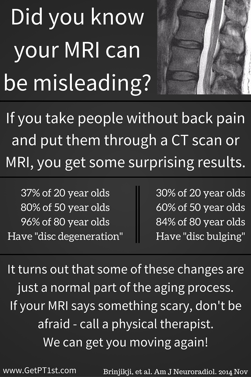
The treatment between the two issues, discogenic back pain and stenotic back pain, is very different. A thorough evaluation can start to correlate symptoms with either discogenic pain or non-discogenic pain. Many patients believe that an MRI will be the answer to why they have pain, but unfortunately this isn’t so.


  1. “a valid and reliable clinical assessment protocol for identifying LSS would be valuable in terms of choosing relevant treatment and informing the patient about the prognosis as early as possible.”


This article was written in 2009. The medical profession has existed for eons. There is still not a valid way to assess a patient in order to determine spinal stenosis. There are biologically plausible ways, meaning that when I assess you, I can make an educated guess from some of the findings in the history and physical, but it is not a valid (proven) way of coming to a conclusion.


  1. “The high sensitivity and specificity of MRI suggests this is a good test for ruling in and out the disease.”


The MRI does a great job of telling us what is abnormal, but it doesn’t do a great job of telling us if the abnormal finding is causing symptoms. As seen in the link above, there are abnormal findings in a population without symptoms. We have to take the imaging findings and see if they make sense after performing a physical exam.


  1. “…history will provide strong clues to the presence of spinal stenosis…more than 65 years of age…prolonged history of low back pain and intermittent radiating symptoms having developed gradually…limited walking capacity…Movements or positions involving flexion e.g. sitting or stooping, will often abolish symptoms…total loss of lumbar extension range is usually found, while flexion most often is well preserved.”


The typical patient with lumbar spinal stenosis will notice that the ability to walk has gradually reduced over time and there is a need to sit due to back or leg pain. Sitting will typically turn down or off the symptoms rapidly. This patient will have limited motion into extension (think of looking over your head to see the stars or bending backwards while standing).


  1. “…stenosis from zygapophyseal joint hypertrophy, ligament thickening or other degenerative changes, it cannot be expected that physical exercise or manual treatment will create a lasting change in the degree of space reduction in the spinal canal or intervertebral foramina”


In the presence of physical changes to the bones, ligaments or loss of disc height, there is nothing that a PT can do to change these back to the way that they were previously. These have been described as wrinkles on the inside. If we look at your face we can start to see how much age you have based on the wrinkles in the face. This is also done on the inside in that some “degenerative” changes are normal. Wrinkles are normal; they are not symptoms of anything sinister. The same can be said for physical changes on the inside. They don’t have to be pain generators. It takes a physical exam to determine how your symptoms respond and whether or not this matches the images on an MRI or X-ray. Even then, we can’t say that movement won’t help, only that we won’t change the physical “inside wrinkles”.


  1. “The main purpose of this pilot study is to evaluate the validity and intertester reliability of an algorithm of physical examination tests, in relation to identifying symptomatic lumbar spinal stenosis.”


This is good. A pilot study is like a pilot for a t.v. show. This is done to see if additional episodes should be done. This study will conclude if additional studies on this topic should be done.   What it hopes to find is a reliable (consistent) way of determining validity (actually seeing what the test hopes to see) in testing for lumbar spinal stenosis. A test that is both reliable and valid should be able to test for spinal stenosis regardless of who is performing the test and who is measuring the test.


  1. “Two patients were classified as “LSS” and five patients “Not LSS”, meaning a 29% prevalence of “LSS” Intertester agreement for overall diagnostic conclusion was 100%”


There are so few patients that this study will likely not yield any results that are actionable. The interesting thing is that the examiners agreed 100% of the time. This is not common in the medical field to have 100% agreement on near anything.


  1. “…the algorithm in its present form can not be used as a screening test to rule out LSS, although it may be able to diagnose the condition.”


There were so few people in the study that it is hard for any clinician to put it to use in the clinic. It may be able to diagnose the condition in that it demonstrated a specificity of 1.0, which is really good.


Excerpts taken from:


Lengsoe L, Lyhne S, Melbye M. An algorithm for clinical identification of spinal stenosis-a pilot study of validity and intertester reliability. International J of MDT. 2009;4(2):21-28.


Can’t find the abstract to the study, but it is listed under the author’s CV http://pure.au.dk/portal/en/persons/martin-melbye(ed4ee688-2d9e-4c17-b0b1-44a5b4b59ada)/publications/an-algorithm-for-clinical-identification-of-spinal-stenosis–a-pilot-study-of-validity-and-intertester-reliability(6d714ee0-d910-11de-9e3b-000ea68e967b).html


Not all back pain is back pain

“Findings such as disk height loss and disc bulges are common in individuals without low back pain.”
Disc bulges, degenerative joint disease, spinal stenosis, can all be a result of living with gravity. We have gravity acting as a compression force on us almost 16 hours a day. Anytime that there is a problem, we want to blame something or somebody. Low back pain is an enigma at times. We can draw correlations, we can come up with risk factors, we can even tell you how to treat it sometimes, but what we can’t do is tell you is exactly what causes your back pain. 
“Surprisingly, disc protrusions were associated with a lower risk of subsequent back pain. Nerve root contact and central stenosis had the largest hazard ratios on baseline imaging findings, and they were associated with incident back pain in the expected direction but not statistically significant. Self identified depression was the strongest predictor of subsequent back pain, with a greeter hazard ratio than any imaging findings.”
What should be taken from the above statistics is that mental health plays a role in pain. There are a lot of new studies that are associating catastrophizing and external locus of control with increased pain levels. Work by Nadine Foster demonstrates a screen for patients who will have a difficult time improving with therapy alone. New were books, such as the one by Annie O’Connor and Melissa Kolski (two people with whom I’ve studied at our RIC study group), goes into great detail regarding pain science. Big picture, we can not neglect the patient’s emotional well-being when attempting to treat the patient’s physical complaints.
“Our results indicate that depression is a strong predictor of who will subsequently report low back pain than baseline imaging findings.Subjects with self reported depression at baseline were 2.3 times as likely to have back pain compared with those who do not report depression.” 
There is obviously a psychosocial component to low back pain. The question is… Chicken or the egg. Is a person more likely to be depressed because they have back pain that is not improving? Or is that person more likely to have back pain because they are depressed? I don’t think that there are cause and affect articles in the literature at this point, but there is definitely a high correlation between patients who are depressed and patient who continue to report low back pain.
“In our analysis of baseline data, we concluded that central stenosis, nerve root contact, and disc extrusion were the most important imaging findings related to prior low back pain. Our current analysis indicates that central stenosis, disc extrusion, and root contact may also be risk factors for future low back pain.”
In other words, if you have a major deformity you will probably have pain. This doesn’t mean that you will definitely have pain, it just increases your risk of experiencing symptoms.
The moral of the story is that we cannot deny the brain. The brain has the ability to see pain, and some patients are more susceptible to seeing this pain. Don’t get me wrong, a thorough mechanical evaluation should be performed when a patient has pain, but when this patient is not inclined to respond to mechanical therapy, the patient should be referred to someone that can better handle this patient’s pain.Sometimes, that person will be a behavioral therapist, a psychotherapist, or a clinical psychologist. Physical therapists are not always the go to in order to treat a patient’s pain.
Excerpts from:

Jarvik JG, Haegerty PJ, Boyko EJ. Three-Year Incidence of Low Back Pain in an Initially Asymptomatic Cohort. Spine. 2005;30(13):1541-1548.  

If the shoe fits

  • SHODDY FOOTWEAR

Understanding the difference among shoes with regards to function

Vincent Gutierrez, PT, MPT, cert. MDT

  • OBJECTIVES

1.To briefly relate the history of the modern athletic shoe

2.To explain the differences regarding functionality among dress shoes, classic footwear, minimalist footwear and barefoot.

3.To provide general recommendations of footwear for varying populations.

  • The Shoe
  • The worlds oldest shoe is roughly 6,000 years old and was meant for foot protection. Prior to this it is theorized that all activities took place barefoot1.
  • Through the years
  • First athletic shoes
  • Keds Champions: unchanged since 19172,3
  • Modern (Classic) Shoe
  • Designed with the foot anatomy in mind (i.e. motion control)3,4,5
  • Minimalist shoes
  • In the recent years these shoes have noted increased sales and are advertised to mimic barefoot activities.
  • These shoes offer no support and increase the intrinsic/extrinsic strength of the foot musculature
  • WALKING
  • When compared to barefoot, wearing standard walking shoes increases stride length by 6%14.
  • Heel strike is more pronounced with larger stride length and varum stresses at the knee were found (9%).
  • Impact on medial compartment OA.
  • For every 1% increase in stress, there is 6x greater risk of knee OA
  • “Flat flexible footwear are associated with significant reductions in dynamic knee loads during ambulation, compared to supportive, stable shoes with less flexible soles.”15
  • Running
  • (1980) It was advised to buy a shoe with built in support mechanisms for the arch and cushioned heel7
  • The authors make this recommendation based on the gait cycle and apply the same gait cycle to running.
  • Recent running analysis challenges this basis of running as fast walking
  • To understand this lets talk GFR
  • http://links.lww.com/CSMR/A3
  • By incorporating arch supports, there is a reduction in elastic recoil of the spring ligament and posterior tibialis, thereby reducing force output at the foot intrinsic/extrinsic3
  • Running
  • Minimalist shoes are more economical compared to classic running shoes in that the the runner utilizes less energy to run9
  • The weight of the shoe was controlled for by using ankle weights.
  • Neuropathic foot8
  • Most ulcers occur in forefoot
  • Study compares barefoot walking in patients with DM neuropathy and those without neuropathy
  • Results
  • Pt with neuropathy place more stress on the forefoot when barefoot (2x more) than controls
  • Possibly due to hammer toe formation and a lack of distribution among toes
  • Unable to feel increased stresses at the forefoot resulting in injury under met. Heads.
  • Balance
  • 100 older women (mean=82 y/a) examining usual footwear vs. barefoot on balance6
  • 68% required AD
  • 42% wore walking shoes, 17% sandals, 11% moccasin
  • Subjects with poorest balance (BBS) benefitted most from usual footwear
  • Post CVA subjects demonstrate increased gait speeds when using a classic shoe compared to barefoot or slippers11
  • Wearing dress shoes (>.5 inch heel) resulted in 15% worsening of balance testing compared to barefoot and a 12% worsening when changing from standard shoe to dress shoe. The TUG improved in standard shoes compared to barefoot12.
  • Healthy older adults demonstrate increased postural sway when wearing traditional walking shoes compared to barefoot13
  • Authors postulate due to sensory deprivation due to footwear
  • Pediatric population
  • “Influence of footwear on the prevalence of flat foot”
  • Study of 2300 children between 4 and 13 y/a
  • 1555 used footwear and 745 never wore shoes
  • 9% of shodded children presented with flat foot and only 3% of children without footwear presented with flat foot.
  • Closed toed shoes appeared to inhibit arch formation moreso than sandals/slippers.
  • The authors suggest that children should play barefoot or in sandals/slippers.
  • Recommendations
  • Running:
  • Classic Running shoes influence a RFS, which increases impact loading into the LE and runners sustain 2.5x more injuries (LBP, LE pain) when running with a RFS3,9
  • Barefoot running fosters a FFS, which strengthens the muscles of the foot3,10
  • Balance:
  • Those with poor balance are advised to wear shoes6 and avoid higher heeled shoes12
  • Healthy individuals are advised to wear minimalist shoes for static balance
  • Neuropathic foot
  • Therapeutic shoes to reduce plantar pressure at the metatarsal heads
  • Walking
  • s/p CVA should wear classic shoes for improved gait speed.
  • Healthy individuals are advised to wear minimalist/barefoot shoes to decrease risk of knee OA
  • Kids
  • barefoot or minimalist shoes
  • QUESTIONS:
  • What’s the difference between running barefoot and running in standard/classic shoes?
  • What are two benefits and limitations of classic shoes?
  • Did this presentation add to your knowledge base and is there a change in your confidence level when recommending shoes for patients/friends?
  • References

1.Ravilious, K. National Geographic News. June 2010. Available at: http://news.nationalgeographic.com/news/2010/06/100609-worlds-oldest-leather-shoe-armenia-science/. Accessed on July 10, 2013.

2.Keds Shoes Official Site. July 2013. Available at: http://www.keds.com/store/SiteController/keds/ourstorypage. Accessed on July 10, 2013.

3.Altman AR, Davis IS. Barefoot Running: Biomechanics and Implications for Running Injuries. Curr Sports Med Reports. 2012;11(5): 244-250.

4.Griffith I. Choosing Running Shoes: The Evidence Behind the Recommendations. February 2011. Available at: http://sportspodiatryinfo.wordpress.com/2011/02/02/choosing-running-shoes-the-evidence-behind-the-recommendations/. Accessed on July 10, 2013.

5.McPoil TG. Footwear. Phys Ther. 1988;68: 1857-1865.

6.Hrogan NF, Crehan F, Bartlett E, et al. The effects of usual footwear on balance amonsgst elderly women attending a day hospital. Age and Ageing. 2009;38:62-67.

7.Heckman B. Selection of a Running Shoe: If the Shoe Fits-Run. JOSPT. 1980;2(2):65-68.

  1. Mueller MJ, Zou D, Bohnert KL, et al. Plantar Stresses on the Neuropathic Foot During Barefoot Walking. Phys Ther. 2008;88:1375-1384.
  2. Perl DP, Daoud AI, Lieberman DE. Effects of Footwear and Strike Type on Running Economy. Med Sci Sports Exer. 2012;44(7):1335-1343.
  3. Lieberman DE. What We can Learn About Running from Barefoot Running: An Evolutionary Medical Perspective. Exerc Sport Sci Rev. 2012;40(2):63-72.
  4. Ng H, McGinley JL, Jolley D, et al. Effects of footwear on gait and balance in people recovering from stroke. http://ageing.oxfordjournals.org/. Accessed on July 6, 2013.
  5. Arnadottir SA, Mercer VS. Effects of footwear on Measurements of Balance and Gait in Women Between the Ages of 65 and 93 Years. Phys Ther. 2000;80:17-27.
  6. Brenton-Rule A, Bassett S, Walsh A, Rome K. The evaluation of walking footwear on postural stability in healthy older adults: An exploratory study. Clinical Biomechanics. 2011;26:885-887.
  7. Keenan GS, Franz JR, Dicharry J, et al. Lower limb joint kinetics in walking: The role of industry recommended footwear. Gait and Posture. 2011;33:350-355.
  8. Shakoor N, Sengupta M, Foucher K, et al. The effects of Common Footwear on Joint Loading in Osteoarthritis of the knee. Arthritis Care Res. 2010;62(7):917-923.

Directional preference compared to “evidenced based practice”

The Audrey Long article is commonly cited as one of the landmark articles for those of us that treat spines utilizing directional preference and centralization.  As well it should be! The results are unbelievable.  This researcher…I take that back…clinician performing research in the clinic published an article that, up until this time, was only speculation.  How could patients not get better using evidence based practice?  For a long time, the evidence was based on expert opinion and not really research.  I enjoy reading the publications on spine and it is interesting to read the changes in the Clinical Practice Guidelines for Low Back, published in JOSPT over the years.  Prior to the most recent publication, flexion based exercises were the rage and directional preference was only moderately supported.  Thanks to clinicians such as Audrey Long, this type of treatment has gained more support in the practice guidelines.  When I was in PT school (I sound like an old man, and year to year this is true, but not yet) this was a very small talking point in our curriculum.  Students now come out of school with a better awareness, though not a true understanding, of the concept of directional preference.


A Critical Appraisal of Directional Preference Exercises Compared to Two Other Exercise Paradigms


P: For patients with low back pain, with or without leg pain, demonstrating a directional preference

I: is treatment with a directional preference

C: as compared to treatment in the opposite direction of the directional preference or an evidence based approach

O: more beneficial when compared with subjective outcome measures


Reviewer:

Vincent Gutierrez, PT, MPT, cert. MDT


Search:

Pedro.org with the keyword terms “directional preference and low back pain”. Nine results were found and the article with the highest score was chosen.


Date of Search: February 15,2014


Citation:

Long A, Donelson R, Fung T. Does it Matter Which Exercise? A Randomized Control Trial of Exercise for Low Back Pain. Spine 2004;29(23):2593-2602.


Summary:


Eleven clinics, from five separate countries, participated in the study. Consecutive patients presenting for treatment of LBP, with or without leg pain, were asked to participate in the study. The inclusion criteria is as follows: consecutive patients with low back pain, with or without one neurological sign, age 18-65 years and demonstrating a directional preference. The exclusion criteria is as follows: cauda equina, two or more neurological signs, spinal fractures, post surgical, off work for one year or more due to low back pain, medical causes, uncontrolled medical conditions, pregnancy, inability to read English (except for those from Germany), patients with prior knowledge of, or specific physician referral for, the Mckenzie method, or no directional preference (DP) elicited.

Therapists credentialed or diplomaed in the McKenzie method performed the assessments. The directional preference was noted as either extension, flexion or lateral, but the subjects were shielded from the significance of directional preference. The subjects were then randomized to one of three groups: matched directional preference exercises, opposite directional preference (ODP) exercises, or evidence-based care (EBC). There were no baseline differences among the groups regarding demographics

The subjects attended at least three and no more than six sessions over the course of two weeks. Those in the DP group received exercises that matched the DP and were instructed to avoid all activities that increase intensity or radiation of symptoms. Those in the ODP group received exercises that were opposite to the DP, and the EBC group performed mid-range exerices and stretches for the hips and thighs. The final two groups also were instructed to return to remain active.


The outcome measures utilized in the study are as follows: back and leg pain intensity ratings using an 11 point visual analogue scale, the Roland Morris Disability Questionnaire (RMDQ), and medication use.


Five hundred three subjects were assessed and 230 demonstrated a DP as follows: 83% extension, 7% flexion and 10% lateral. Twenty-nine dropped out of the study at two weeks, and the remaining 201 were eligible for analysis. There were 36 withdrawals, which indicated that the subjects worsened or had no change in symptoms and were transitioned to alternative care. None of the DP group withdrew, but 16 in the ODP and 20 in the EBC withdrew. All outcome measures improved in the three groups over the course of two weeks, with the DP group demonstrating significant improvement compared to the ODP and EBC.


Appraisal:

The authors satisfied eight of the ten questions regarding the Quality Appraisal Checklist. The subjects’ group design was not blinded to those enrolling the subjects and this was a comparison study of varying interventions, which indicates that a true control group was absent.


This study will have a direct impact for clinical therapists. Because this study compared three different interventions, opposed to identifying the efficacy of a single intervention compared to a control group, it mimicked clinical practice. The authors compared evidence based care with a directional preference treatment paradigm, which would be similar to a question asked in clinical practice.


Conclusion:

Directional preference exercises are superior to performing exercises opposite to the directional preference or “evidence based care”. Patients that demonstrate a directional preference and are treated accordingly perform significantly better in outcomes measured in this study. There appears to be no harm in treating a patient with directional preference exercises, but the same does not hold true for performing exercises opposite of the directional preference or “evidence based care”.

The following is breakdown of the systems involved in “assessing” research articles.


  1. Were the subjects randomly assigned into groups?

Yes. The subjects were randomly assigned to one of three groups.


  1. Was each subject’s group assignment concealed from the people enrolling individuals in the study?

No. Because the study is a multi-centered study in outpatient practice, it was not acceptable to the authors to have patients drop out of the study due to changing therapists.


  1. Did groups have similar characteristics at the start of the study?

Yes. The authors note that there were “no differences among the three treatment groups in any baseline demographic characteristics or outcome measures.”


  1. Were subjects masked or blinded to their group assignment?

Yes. Although subjects couldn’t be blinded to the treatment, they were unaware of the specific grouping (i.e. matched vs unmatched vs EBP)


  1. Were clinicians and/or outcome assessors blinded to the subjects group assignment?

No and yes. Although the clinicians were not blinded, which is common in practice, the assessors of the outcome measures were blinded. The outcome measures were subjective measurements in order to minimize therapist bias.


  1. Did the investigators manage all of the groups in the same way except for the experimental investigation?

No. Because this is an intervention study in patient’s seeking treatment, it was impossible to withhold treatment to establish a true control group. The types of treatment for the opposite direction and EBP groups were vaguely described.


  1. Did the investigators apply the study protocol and collect follow-up data on all subjects over a time frame long enough for the outcomes of interest to occur?

No. There was a 12% dropout rate, which was anticipated by the authors when determining the number of patients needed to maintain a power of .90. Thirty-eight patients withdrew from the study, as opposed to dropped out, due to no improvement or worsening of symptoms in the EBP and unmatched group. Two weeks was long enough in order to assess change.


  1. Did subject attrition occur over the course of the study?

Yes. Twenty-nine subjects dropped out of the study, with 12 of the 29 dropping out due to no change or worsening of symptoms. Although these participants did not “drop out”, indicating that follow-up information was unattainable, 36 subjects withdrew early due to no change or worsening of symptoms. No subjects from the matched group withdrew. Those that withdrew completed the outcome measures prior to 2 weeks in order to seek alternative care.

MRI: Medico-Reckon to identify: what you need to know


MRI: Medico-Reckon to identify: what you need to know.


This was a great article. It puts numbers to the faces seen on MRI’s. I like numbers…kind of like Rainman. Numbers comfort me. Enjoy the read. There is some higher level thinking in the below quotes. If you have any questions, leave a post either here or on the movementthinker Facebook page.


  1. “Magnetic resonance imaging (MRI) provides clinicians with a noninvasive mechanism for viewing lumbar anatomy in great detail”


READ AND RE-READ THE ABOVE STATEMENT.

Question #1 from the above statement: Can an MRI tell me what is causing your pain?


Question #2: Can an MRI tell me how to treat you?


Question #3. Does the MRI differentiate between abnormal structures that cause pain and abnormal structures that don’t cause pain?


The answer to all of the above questions is NO! Everyone seems to think that they need an MRI before they come to therapy…as if I am going to just treat them on a whim without the MRI…or that the MRI will somehow give me a paint by number way of treating the symptoms. This does not exist. The MRI can be helpful in a small percentage of patients that are either seeking or needing surgery, but aside from that it is just something for me to read after I have performed my clinical assessment of the patient and come up with my own conclusion. Now…if my conclusion matches the MRI then awesome! Well…at least for me. If it doesn’t match the MRI…that sucks because now I have to go back and reassess to see which one of us is more right…the PT or the MRI.


  1. “For example, large variations in lumbar disc and radicular canal morphology have been identified in both symptomatic and asymptomatic individuals”


This means that an MRI is very good at determining what is not normal, as compared to a textbook, but the variations of normal is so wide that the test may not tell us much.


  1. “…challenge for examiners in their attempts to differentiate between observations that are “symptom generators” and those that are benign variations”.


When a radiologist reads your MRI, they are the ones that are determining what is going on in the pictures, they spend on average of 30 seconds per picture. In 30 seconds, they have to figure out what is abnormal. Then, if they have found something abnormal, they have to determine if it can cause your symptoms. All of this is performed without ever evaluating the most important aspect of the symptoms…YOU! The radiologist never sees you. If you look at the bottom of your report (assuming that you have already had an MRI), you will typically see the phrase “patient would benefit from clinical examination to correlate imaging”. This is the radiologist saying; “Look, I only have the pictures. I can tell you with a degree of certainty what the MRI says…does this fit your symptoms?”


  1. Patients were classified according to this table:


  1. Primay LBP (low back pain): pain in the back or buttock


  1. Posterior thigh referral: pain in one or both back/lateral thighs with or without LBP


  1. L1-L3 distribution: pain in the anterior thigh and top of foot


  1. L4-5 distribution: pain in the mid and distal anterior thigh, anterior leg and top of the foot.


  1. S1-S2 distribution: Pain in the lateral border of the foot and bottom of the foot


  1. Bilateral distribution: any combination of the above in both legs instead of one leg.


  1. Atypical: none of the above.


This is an overall pain pattern distribution. Unfortunately, this is not drilled in PT school. I was about 2-3 years out before I figured this out on my own and then after discovering it, I looked it up. It’s funny…if you don’t know what you don’t know, then you don’t know how to find it. I think that PT’s schools should heavily bias students in this direction for learning. Think of it. If you knew that for every dollar you invested, you would get an 80% return if you simply knew a few tricks…would you learn those tricks?


Roughly 80% of the population will have back pain at some point in his/her life. This is either the primary or secondary reason for physician office visits (depending on which research you read) and the one that it competes with is the common cold. Think about that…back pain is about as “common” as the cold.


  1. “All images were initially screened for evidence of neoplastic, inflammatory or infectious disorders…”


This is all of the very bad stuff that needs to be ruled out if someone is going to look at an MRI. This is stuff that won’t get better with therapy. If you have certain characteristics, your PT may refer you back to your physician in order to rule out the nasty stuff.


  1. “…study involved 408 participants…55% had acute pain…50 participants reported a recurrence of previous symptoms within the past 2 months…303 participants reported chronic symptoms of longer duration than 2 months”


This sounds about right. Those with back pain may have it go away, but it will come back. Those whose pain doesn’t come back is mostly because…IT NEVER WENT AWAY!


  1. “…the most common location of symptoms was in the S1-S2 segmental, followed by the L4-L5 distribution. Bilateral radicular patterns were the least frequent.”


This means that a high percentage of patients had symptoms radiating into the foot, from the back. Fewer patients experienced symptoms into both legs. If both legs are causing you pain…at the same time…you are among the few.


  1. “The presence of weakness in ore of both lower extremities was reported by 175 participants (42.9%)”


If your back symptoms are bad enough, they will start to cause a “power outage”.   For instance, I use a specific analogy in the clinic. If your lamp doesn’t turn on when you flip the switch…what is wrong?   A common answer is that the light bulb is burned out. How many light bulbs will you go through before you realize that the bulb is working fine? When a muscle is weak, it is like the above idea. I can give you strengthening, but I would have to give you about 6 weeks of strengthening exercises in order to determine if “just muscle weakness” is the problem. This is like changing the light bulb daily for 6 weeks. I doubt that you would actually do this. Most people may do this once or twice and then just give up. When I give you strengthening exercises, you will do them for a couple of days and then give up because you won’t see much change.


What else could cause the light to not turn on? There could be a fray in the cord. This also happens in the body. If there is a nerve (electrical wire) that is not working appropriately, then the muscle won’t contract…the light bulb won’t turn on. This one becomes a little harder to figure out because we would have to try to find the location of the “fray”.


The final thing is the easiest to check for…the lamp isn’t plugged in.


It’s funny because I frequently have students. Recently, I had a patient that struggled to go up the stairs. She noted that her leg was weak. Students always want to make a muscle stronger. They are good at that. Unfortunately, her hip muscle wasn’t plugged in. After performing 30 repetitions of repeated extension in lying, her hip strength went from weak to moderately strong. Her ability to ascend stairs was visibly improved and the patient was surprised that her sensation of strength had improved. The student asked “why don’t we learn this in school?” I don’t know. I have the same question.


  1. “Disc extrusion was significantly related to the presence of distal lower extremity pain…not significantly related to weakness…not significantly associated with the presence of paresthesias or numbness”


What is a disc extrusion? This guy does a great job of explaining it: http://www.bodiempowerment.com/disc-bulge-why-is-my-disc-bulging/

Why reinvent the wheel?


  1. “Overall 149 of the participants (37%) had MRI evidence showing some degree of nerve or thecal sac compression…The most common segmental level of compression was L4-L5, followed by L5-S1…There was a significant association between the side of nerve compression and the side of pain…of the 256 patients with no evidence of nerve compression visible on MRI, 151 (58%) indicated unilateral lower extremity symptoms”


This means that some patients that have an MRI will show that the disc has caused some sort of nerve compression. When this happens, you will typically have pain on the side of the compressed nerve. On the flip side though, you can have pain on in one leg that is not coming from the nerve. Think like this…nerve compression can cause leg pain, but not all leg pain is caused by nerve compression.


  1. “participants who reported weakness had a greater prevalence of nerve compression, and those without weakness had a lower prevalence of nerve compression”


Again, the nerve supplies electricity to the light bulb. If the electricity is not getting there because of a problem with either the plug or the cord, then the muscle won’t work.


  1. “Roughly 63% of the participants had no evidence of nerve root compression on MRI. Of these, 35% had pain patterns referring distally to the knee”


THIS IS HUGE! PT’s in school learn that if you have pain below the knee that there must be some nerve that is compressed. This is not always the case. Any structure that has a nerve going to it can cause pain to radiate in a pattern specific to that nerve. For instance, in the neck we know that if we irritate the nerve in the joint, it could refer pain into the shoulder blade. It doesn’t have to be a “PINCHED NERVE”!


  1. “the presence of disc extrusion or ipsilateral, severe nerve compression at one or multiple sites is strongly associated with distal leg pain. Mild to moderate nerve compression, disc degeneration or bulging and spinal stenosis are not significantly associated with specific pain patterns.”


I enjoy weightlifting. When I see a snatch done well, it is like poetry. I can’t explain the entire movement in one fell swoop other than to say it is beautiful. When I see someone do this movement, with little experience, we can officially say that: yes you went from point A to point B, but not well.


When we see a sever nerve compression or disc extrusion, we can say “YUP I KNOW WHAT THAT IS.” Anything past that is a guess as to what is causing your symptoms, based on the MRI.


Quotes taken from the following:


Beattie PF, Meyers SP, Stratford P et al. Associations Between Patient Report of Symptoms and Anatomic Impairment Visible on Lumbar Magnetic Resonance Imaging. Spine 2000;25:819-828.


You are not your MRI..at least not for long

did-you-know-your-mri-can-be-misleadingYou are not your MRI…at least not for long.


MORAL OF THE STORY: Stop your whining over your herniated disc, bulging disc or exploding disc. You are probably not the outlier. If your pain is lasting longer than six months, your disc is probably healed, but you still move like crap. Start to move better and take better care of yourself and the improvements will follow. In general, this means that you are most likely the problem…not your back.

Also, I will be taking a couple of weeks off from reading and writing to travel with the family.  Taking some time to breathe.  If you enjoy the blog, please add a topic that you would like to see covered at a later date.


  1. “Lumbar disc hernia (LDH) is a common cause of low back pain and radicular leg pain…majority of LDH patients recover spontaneously…Purpose of the present study was to investigate the natural history of the morphologic changes of LDH on MRI and to assess correlations with the type of LDH and the clinical outcome”


First, disc herniations are a common cause of pain. I believe this to be true and the research consistently reports this fact. The part that doesn’t get reported is the second part of the statement being that spontaneous recovery is normal.


When people come into the clinic, they have this seemingly rehearsed story of how they had an MRI and was told that they have a bulging/herniated/exploding (maybe a little overboard) disc. The doctors never tell them that this can recover on its own and patients then wear the herniated disc patch for the rest of their lives.


As you will see, you no longer need to wear that patch if your were told that you have an exploding disc.


  1. “…42 patients…mean age of 42…unilateral leg pain and low back pain…symptomatic level was L2-3 in 8 cases, L3-4 in 6 cases, L4-5 in 15 cases and L5-S1 in 13 cases”


Let’s start here. l_spine


The lumbar spine is labeled as L1-5 and the sacral spine then starts. The intersection between the lumbar spine and the sacrum is L5-S1. The segments are named by the upper segment first-lower segment second.


Some interesting notes regarding this study:


  1. 66% of the patients have symptoms coming from the lower lumbar segments, those being L4-S1. This is inconsistent with published research reporting that up to 95% of symptoms come from these lower segments.
  2. Therefore, 34% of symptoms are coming from the upper segments. Again, previous research notes that only 5% of symptoms come from these segments.


Unilateral leg pain simply means that only one leg is affected. For those that may have experienced sciatica in the past, you will remember that it was only one leg that experienced symptoms. If you have symptoms in both legs, then it may not be sciatica.


  1. “All patients underwent MRI examinations every three months for a period of 3-24 months”


This is not affordable for most and won’t be approved by any insurance that I have encountered. The reason for the frequent MRI’s is to see how things change over time.


  1. “LDH was classified into three types: protrusion (n=7), extrusion (n=17 and sequestration (n=18)”


Here comes the jelly donut theory. If you have heard it, then you can pass this paragraph up. Think of the disc as a jelly donut (I know that this is an oversimplification, but this model makes the most sense…even if it is not the most accurate).


A protrusion means that the outer portion of the donut (the actual donut itself) has been deformed. If you plug the hole of the jelly donut so that the jelly can’t come out of the hole, you will be able to follow along with the rest of the idea. I personally don’t like jelly donuts. I much prefer custard or cream. Speaking of that, Tim Hortons has the best filled donuts that I have ever had. This reminds me of a trip to Canada with my best buddy Carl. If I have the time later, some stories from this road trip may come out. Back to business; if you squeeze the donut on an edge lightly, you will start to squeeze the jelly away from the area that you are squeezing. If you squeeze a little harder, you will see the donut “bulge” just prior to the jelly coming out. This is a protrusion.


An extrusion means that the jelly has escaped! Oh no! Now what? No big deal. You will see later that this may actually be a better situation for you than the protrusion.


A sequestration means that not only has the jelly escaped, but a piece has broken off and hit the floor. If enough nuclear material (the jelly inside the disc) breaks through the annulus (the donut in the example), then it may break off and be free floating in the spinal canal (near the nerves of the spine). This again may not be as bad as it sounds.


  1. Correlation between the clinical outcome and spontaneous changes of the herniated mass on MRI (6 months)


MRI change Excellent Good Poor Total%
Disappearance 6 2 0 19
More than 50% reduction 11 18 0 69
Little or no reduction 0 1 4 12
Total 40 50 10 100


What this means is that in 19% of patients, the herniation seen on the MRI disappeared over the course of time. Better yet, about 88% improved significantly over the course of time. You are not your MRI… at least not for long.


6.

Type of herniation Case Duration of symptoms
Protrusion 3 cases in total 3-14 weeks with 8 weeks average
Extrusion 17 cases 4-8 weeks with 4.8 weeks average
Sequestration 18 cases 1-5 weeks with 3.2 weeks average


What does this chart mean? Those that have a “more serious” appearing herniation on MRI actually respond faster than those with a smaller herniation. You are not your MRI…at least not for long


Excerpts taken from:


Takada E, Takahashi M. Natural history of lumbar disc hernia with radicular leg pain: Spontaneous MRI changes of the herniated mass and correlation with clinical outcome. Journal of Orthopaedic Surgery. 2001;9(1):1-7.

If it hurts it must be bad, or good, or whatever. Vincent Gutierrez, PT, cert. MDT

Louw A, Puentedura EJ, Zimney K, Schmidt S. Know Pain, Know Gain? A perspective on Pain Neuroscience Education in Physical Therapy. J Orthop Sports Phys Ther. 2016;46(3):131-134.


  1. “Pain is a normal human experience and essential to survival”

This portion is rarely spoken of in PT school and we spend our time in school learning how to shut down the pain, either in an ideal way of dealing with a mechanical problem or in a way in which we “trick” the brain of not seeing the pain for a short period of time. When working with patients, I often describe the gate control theory as the “Three Stooges” way of treating pain. For instance, if you have a headache and I hit your foot with a hammer, what happened to your headache. I stole the example from my dad, because this is how he would always respond if I told him my arm was sore after baseball practice. This was way back in the 1980’s and he was a laborer by trade. The gate control theory makes sense to most people, but we can also see the example and understand that it is probably not the best way to fix a problem, as we end up with a broken foot from the hammer.


  1. “The pain neuromatrix explained our knowledge and understanding of the functional and structural changes in the brains of people suffering from chronic pain”

To simplify, we have pain because our brains tell us that something is painful. This could be due to past experiences, actual painful stimuli eliciting Nociception, super excited nerves , so on and so forth.


  1. “biomedical models may induce fear and anxiety, which may further fuel fear avoidance and pain catastrophization”

It is very common for a patient to come into the clinic and say that he/she is avoiding a particular activity because of a history of a herniated disc. There is research that shows that a herniated disc can become “unherniated” (for a lack of a more layman’s term) over the course of 6 months. The patients are never educated regarding this point. Once a herniation, always a herniation is just not true. This biomedical or pathoanatomical (patho=bad and anatomical = body parts) model of health care is outdated and simply is not as useful to use with the general public because research demonstrates that the patient may become “sick listed” and from there stop participating in previously enjoyable activities.


  1. “a plethora of papers have been dedicated to a mere 20-millisecond delay of abdominal muscle contraction, yet despite the enormous amount of time, money and energy spent on this science, clinically it has yet to provide results superior to those of any other form of exercise for low back pain”

Doing the vacuum pose while lying down is no better than doing a general squat or learning how to utilize your diaphragm during breathing mechanics. As the layperson, there are many people that want to take your money in the health care industry. (I hate to say it like this, but healthcare is a huge business and the public needs to see it as so.) When the new fad comes out to solve back pain, don’t buy into the infomercial and as a matter of fact, turn off the t.v. and go get a book from the local library. You will spend hundreds of dollars less than what is proposed on the infomercial and be better off after having read the book. Nothing beats knowledge and the smarter you are at taking care of yourself, the better armed you are when you actually get in front of a health care practitioner. Remember, it is a business and we all want your money if you will give it to us. A better use of your time is to come educated so that I don’t have to teach you the basics of posture for 30 minutes, but can instead can teach you how to perform more high level movement patterns instead of sitting properly to reduce your pain. Oh wait, pain is normal. I’d lose my job if I sold this to all of my patients, but instead the patients need to be educated between hurt and harm.


  1. “In all health care education, be it smoking cessation, weight loss, or breaking addiction, the ultimate goal is behavior change.”

Speaking as a physical therapist, I can’t stress to the patients enough how the therapy experience is a team. Smart people call it therapeutic alliance, but I’ll settle for team. My part is to educate the patient and attempt to solve the puzzle of the patient’s pain, but it is the patient’s job to take the information that they have gained during the session and go home and apply it to their daily lives. For a patient to do nothing at home, AKA make no changes in behavior, and come to the following session thinking that the pain will go away is similar to :


https://spencergarnold.files.wordpress.com/2013/01/snatch-miracle.jpg


Patients may come hoping for a miracle, but it is not to be. The patient and therapist have to work together to attempt to solve the pain problem. If one side of the team is not doing their part, then the PT has to be willing to discharge the patient or the patient has to be willing to fire the PT.


  1. “…when PNE (Pain Nueroscience Education [pain is a normal human response]) is paired combined with either exercise or manual therapy, it is far superior in reducing pain compared to education alone”

From this I take that teaching the patient and then moving the patient is better than just teaching the patient. We can all agree that low level exercise is good for people. If we don’t agree with this, then we are saying that it is safer long term to live like a slug then to get up and walk around the living room. It just isn’t so. People will refuse to get up and walk around the living room when they start experiencing low back tightness, leg fatigue, or the dreaded “Fran cough” (look it up and btw I am an advocate professionally speaking). We as a society have to start moving more and learn about how our body is supposed to work. This can not be done from infomercials that have pictures of pulsating backs or frowning stomach fat.

And this is my two cents for the night.
If you are in need of physical therapy or would like to sign up for a complementary discovery session (a conversation to determine if therapy is right for you), contact me. 

Functional Therapy and Rehabilitation 

(Now part of the Goodlife family)

903 N 129th Infantry Dr. 

Joliet Il 60435

815-483-2440

Rehab post TKA

Piva SR, Gil AB, Almeida GJM, et al. A Balance Exercise Program Appears to Improve Function for Patients With Total Knee Arthroplasty: A Randomized Clinical Trial. Phys Ther. 2010;90:880-894.

Intro: 37% of TKA’s still have functional limitations p one year. Diminished walking speed, difficulty ascending/descending stairs, inability to return to sport are chief functional complaints. During TKA surgery several tendons, capsule, and remaining ligaments are retightened to restore the joint spaces deteriorated by the arthritis. Some of the knee ligaments are removed or released, which may affect mechanoreceptors/balance.

PURPOSE:

  1. To determine the feasibility of applying a balance exercise program in patients with TKA
  2. To investigate whether an F (functional) T (training) program supplemented with a balance exercise program (FT+B) could improve function compared to FT program alone
  3. To test the method and calculate a sample size for a future RCT with a larger sample size

METHOD: Double-blind pilot RCT (very strong evidence)

Inclusion: TKA in the previous 2-6 months (meaning not eligible for study if the TKA was before 2 months previous)

Exclusion: 2 or more falls in the previous year. Unable to ambulate 100 feet with an AD or rest period, acute illness or cardiac issues, uncontrolled HTN, severe visual impairment, LE amputation, progressive neurological disorder or pregnant (interesting exclusion criteria).

All went through a quadriceps muscle-sparing incision (cuts through the fascia of the patella instead of the quadriceps) this may be a factor in reducing rehab stay.

See the appendix for the protocol (6 weeks).

Testing measures:

  1. Self-selected gait speed (interesting, but probably not feasible for our clinic)
  2. Timed chair rise test (5 repetitions): easily added to our testing.
  3. single leg stance time: easily added in
  4. LEFS
  5. WOMAC

RESULTS:

  1. Adherence for both groups is 100% and the HEP adherence was similar (filled out logs)
  2. walking speed continued to improve over the course of 6 months for the FT+B group and was 25% better than the FT only group.
  3. The interesting fact is that improvement continued up to 6 months, when previous literature describes 3 months and done.
  4. Single leg stance: FT+B improved (as expected due to SAID), but the FT group either maintained or worsened on speed and balance.

DISCUSSION: FT+B demonstrates clinically important differences in walking speed, SLS, stiffness and pain, without adverse events. Subjects in the FT+B could balance on average 4 seconds longer than baseline. This may be important for weight bearing during the stance phase of walking. Performance-based measures should be used in place of subjective measures.

TAKE HOME: Patients will benefit from the addition of balance exercises post-surgically. It may be prudent to discuss with the surgeons of increasing the length of stay in therapy and decreasing the number of visits per week, as progress continues to occur past the 3 months initially surmised. Each patient should be tested with one or more of the following:

  1. SLS
  2. Chair rise test
  3. Gait speed: important indicator of function/independence/death
  4. Balance test (excluding Tinetti due to possible ceiling affect when the patient no longer needs an AD).

If you need therapy after a total knee replacement, you can contact me at the following location.
Dr. Vince Gutierrez, PT, cert. MDT

Functional Therapy and Rehabilitation (Now part of the Goodlife family)

903 N Infantry Dr.

suite 500

Joliet, IL

60435

815-483-2440